
They may also be used to see how the teeth touch each other when the patient bites down.

The main use of a bite-wing x-ray is to see if there is tooth decay. They can also show what’s underneath existing fillings. The images will show the biting surface and the area where the main part of the tooth meets the root. They are not typically done on front (anterior) teeth. The patient bites down on the tab so the image will show both top and bottom teeth.ĭentists use bite-wings to get a picture of the back (posterior) teeth. They get their name from a tab on the x-ray film. There are four common types of dental x-rays, each used for different purposesīite-wing x-rays are the type that most people are familiar with. Cavities, infections, and other conditions show up as dark spots on the lighter image of the tooth.ĭifferent types of x-rays have different purposes, depending on what the dentist is trying to see. It is one of the dentist’s most important diagnostic tools, giving him or her a better picture of what’s going on with your teeth than simply looking in your mouth.ĭental radiographs work by using a small, controlled burst of radiation to create a picture of the tooth. A dental x-ray is the common term for a dental radiograph. In addition to a thorough examination of the mouth and teeth during these appointments, the dentist will sometimes order x-rays. Before booking an appointment with us please ensure that you have already a sufficient referral from your practitioner with the justification for exposure, otherwise please contact our surgery and ask for a referral form.Good dental health should include regular visits to the dentist.

Private Medical Centre in Ealing welcomes all patients requiring an OPG. Bite wing program for the anterior tooth region and left/right side.All artifacts reduced with constant magnification - for patients with numerous fillings.Panorama with constant magnification 1.25.Standard panorama half-sides: left or right, upper or lower jaw.Standard panorama with orthoradial beam direction- for the basic diagnosis.With Orthophos XG 3D-ready our dental practice offers: individual adaptation of the imaging layer to the anatomy of the patient, i.e.lower the exposure level making it available for children or even pregnant woman.explain treatments to patients with greater clarity and accuracy.coordinate planning treatment with colleagues.
#PANORAMA X RAY SOFTWARE#
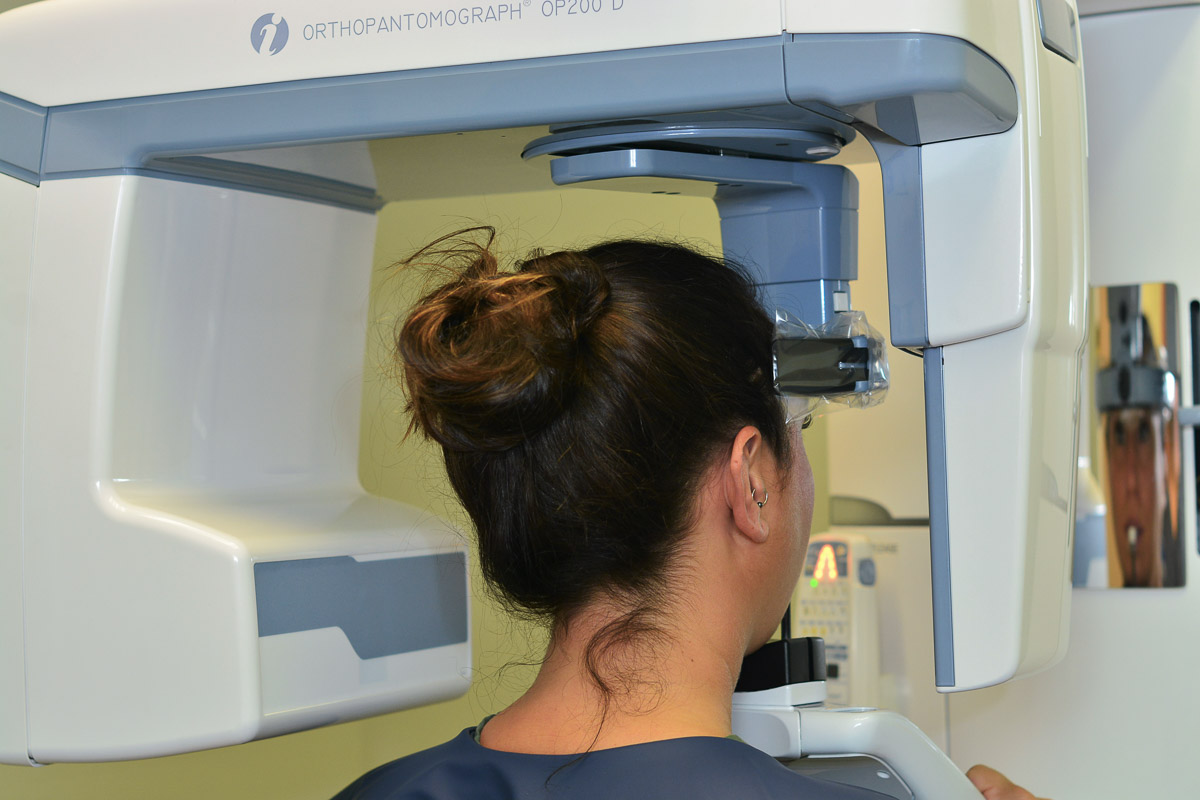
Digital technology captured inside ORTHOPHOS XG 3D-ready extends dental diagnostic X-ray imaging potential in the fields of:Īdvanced level of technology and visualization tools available in the software and hardware of Orthophos XG 3D-ready unit enable practitioners to: With Orthophos XG 3D-ready from Sirona (a Germany based World Leader in Dental Technologies) you will see more in dental diagnostics. Best image quality, lowest dose of radiation and perfect workflow are one of the key advantages of this imaging technology. This technology allows capturing patient’s whole jaw in a single scan and shows the two-dimensional or three-dimensional (3D models) view of a half-circle from ear to ear. In Private Medical Centre we bring dental diagnostic X-ray imaging to a higher level with the use of ORTHOPHOS XG 3D-ready – the best and most popular advanced digital X-Ray imaging machine in the World! A panoramic radiograph, also called an OPG, is a result of panoramic scanning dental X-ray of the upper and lower jaw. ORTHOPHOS XG 3D-ready New dimension dentistry


 0 kommentar(er)
